Chest X-ray Masks and Labels
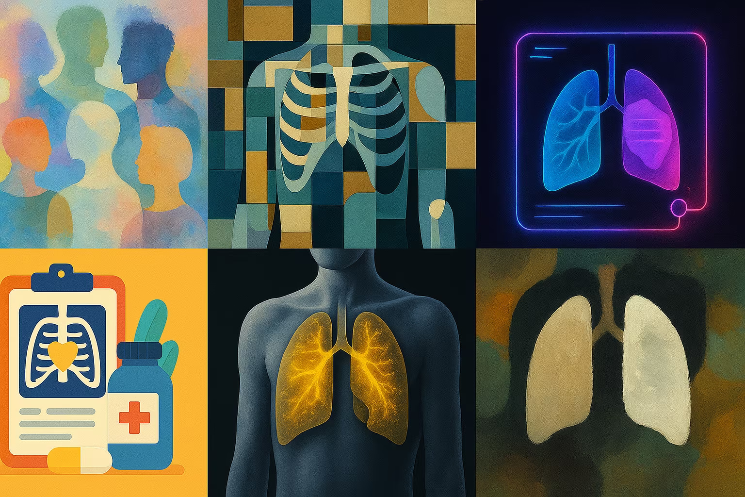
Chest radiographs accompanied by lung segmentation masks, useful for tasks such as the detection of pathologies or domain adaptation.
Description
This dataset provides a set of medical images composed of chest x-rays and associated segmentation masks. The images come from several hospitals and health institutions, with a variety of lung conditions represented, including tuberculosis. Some masks may be missing, which invites manual verification or dataset enrichment.
What is this dataset for?
- Training lung segmentation models on medical x-rays
- Develop classifiers to detect tuberculosis or other lung abnormalities
- Evaluate the robustness of models on images from different clinical sources
Can it be enriched or improved?
Yes, it is possible to complete the missing masks, to further annotate visible anomalies or to adapt the images for multilingual scenarios. Improvements like automatic alignment, intensity normalization, or data augmentation are relevant here.
🔎 In summary
🧠 Recommended for
- Medical imaging researchers
- Lung segmentation projects
- Adaptation of the medical field
🔧 Compatible tools
- MONAI
- PyTorch
- MedPy
- ITK-SNAP
- Keras
💡 Tip
To improve results, use augmentation techniques specific to X-ray imaging such as contrast inversion or Gaussian blur.
Frequently Asked Questions
Are the masks complete for each x-ray?
No, some x-rays do not have an associated mask. It is advisable to check manually or to complete with your own annotations.
Can this dataset be used to train an automatic diagnostic model?
Yes, for research purposes only. It should not be used for clinical purposes without regulated medical validation.
Are the images anonymized and ready to use?
Yes, all images are de-identified and provided in a DICOM format adapted to deep learning processing.